C30C Mini Color Ultrasound Diagnosis System
- PRODUCT DETAIL
C30C Mini Color Ultrasound Diagnosis System
Product Introduction
The
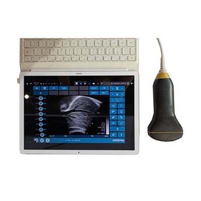
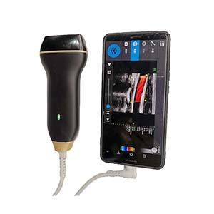
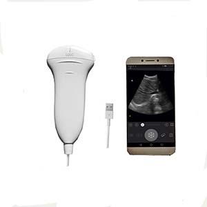
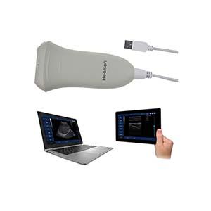
C30C Mini Color Ultrasound Diagnosis System is a portable handheld ultrasound
imaging device designed for rapid clinical examinations, point-of-care
diagnostics, and preliminary screening. Featuring an integrated convex array
probe and USB connectivity, it connects to compatible Windows or Android
terminal devices for convenient real-time ultrasound imaging.
The
C30C supports multiple imaging modes, including B-mode, B/B-mode, B/M-mode,
color Doppler flow imaging (CDFI), pulsed-wave Doppler (PW), and power Doppler
imaging (PDI). Its digital beamforming, dynamic focusing, adjustable frequency,
and advanced image-processing technologies help clinicians visualize abdominal
and pelvic organs, including the liver, gallbladder, kidneys, spleen, pancreas,
uterus, and bladder.
With
a compact probe weighing no more than 150 g, preset examination protocols,
image storage, and playback capabilities, the C30C is suitable for hospitals,
primary healthcare facilities, emergency services, bedside examinations, and
ultrasound-guided procedures. Compatible smart terminals can also support image
sharing and remote medical workflows, depending on the software and
connectivity configuration.
Performance Features
• Compact
Handheld Design: Integrated convex array probe measuring no more than 155 ×
75 × 28 mm and weighing no more than 150 g for convenient transport and
operation.
• Multiple Imaging Modes: Supports at
least six imaging modes: B, B/B, B/M, CDFI, PW, and PDI.
• Advanced Digital
Imaging: Incorporates digital beamforming (DBF), real-time dynamic aperture imaging
(RDA), dynamic sound velocity adjustment (DRA), and dynamic transmit and
receive focusing technologies.
• Wide Frequency
Selection: Offers six selectable frequencies from 2.5 MHz to 5.0 MHz to accommodate
different examination requirements.
• Adjustable
Imaging Depth: Provides a detection depth of 90–240 mm, adjustable
through the connected terminal.
• Preset Examination
Protocols: Includes abdomen, gynecology, obstetrics, and urology examination presets, with
options to customize examination sites.
• Flexible
Image Adjustment: Provides adjustable brightness, contrast,
grayscale curves, dynamic range, frame correlation, noise suppression, and
false-color settings.
• Image Magnification: Supports
eight levels of real-time image magnification, up to 4×, plus partial
magnification for real-time and frozen images.
• Comprehensive
Measurement Tools: Supports distance, circumference, area,
volume, angle, time, slope, heart rate, and Doppler velocity measurements,
depending on the selected imaging mode.
• Portable USB
Connectivity: Connects to compatible terminals through USB Type-C or USB
Type-A interfaces.
• USB-Powered Operation: The probe
does not require a separate battery. The system can operate continuously for at
least 3 hours under specified conditions.
• Image Storage and
Playback: Supports static image formats including BMP, JPG, PNG, TIF, and DICOM (DCM),
plus storage and playback of up to 2,048 frames.
• Clinical Documentation: Offers
patient information entry, annotations, body-position markers, probe-position
indication, and Chinese, English, and French interface options.
• Data Transfer Support: Supports
image transfer to a USB flash drive and DICOM sending on Windows, subject to
compatible software and system configuration.
Working Principle
The
C30C Mini Color Ultrasound Diagnosis System uses electronic convex array
ultrasound technology combined with digital beamforming and advanced signal
processing to generate diagnostic images.
1. Ultrasound
Transmission: The integrated probe transmits high-frequency ultrasound
waves into the body.
2. Echo Reception: Reflected
ultrasound signals from tissues and organs are received by the probe's
transducer elements.
3. Digital Beamforming: Digital
beamforming and dynamic focusing process the received signals to construct
ultrasound images.
4. Image Processing: Dynamic
aperture control, frequency adjustment, digital filtering, frame correlation,
grayscale processing, and image enhancement improve image presentation.
5. Doppler Imaging: CDFI, PW,
and PDI modes provide blood-flow-related information through Doppler-based
signal processing.
6. Display and
Documentation: Images are displayed on a compatible Windows or Android
terminal, where users can perform measurements, freeze or magnify images, save
examination data, and review recorded frames.
The
combination of portable hardware and digital imaging software supports flexible
ultrasound examinations in clinical and emergency environments.
Technical Parameters
Parameter
Specification
Product
Model
C30C
Product
Type
Mini
Color Ultrasound Diagnosis System
Probe
Type
Portable
handheld convex array probe
Scanning
Technology
80-element
electronic convex array
Probe
Dimensions
≤155
× 75 × 28 mm
Probe
Weight
≤150
g
Supported
Operating Systems
Windows,
Android
Display
Modes
B,
B/B, B/M, CDFI, PW, PDI
Operating
Frequency
2.5,
3.0, 3.5, 4.0, 4.5, 5.0 MHz
Detection
Depth
90–240
mm, adjustable
Dynamic
Range
0–100
dB, adjustable
Grayscale
Levels
256
Brightness
Adjustment
16
levels
Contrast
Adjustment
16
levels
Grayscale
Curve Adjustment
13
levels
Frame
Correlation
8
levels
Noise
Suppression
7
levels
False-Color
Options
16
Display
Speed
3
adjustable levels
Image
Magnification
8
levels, up to 4×
TGC
Adjustment
8
segments
Image
Storage
BMP,
JPG, PNG, TIF, DCM and other supported formats
Cine
Memory
Up
to 2,048 frames
Data
Transfer
USB;
DICOM sending supported on Windows
Probe
Interface
USB
Type-C and USB Type-A
Power
Supply
USB-powered
probe; no separate probe battery
Continuous
Operating Time
≥3
hours under specified conditions
Preset
Examination Modes
Abdomen,
gynecology, obstetrics, urology
Interface
Languages
Chinese,
English, French
Application Scenarios
• Abdominal Ultrasound: Examination
of the liver, gallbladder, kidneys, spleen, pancreas, and other abdominal
organs.
• Gynecology and
Obstetrics: Supports pelvic examinations and obstetric ultrasound
workflows, including preliminary fetal assessments where clinically
appropriate.
• Urology: Assists with
ultrasound examinations involving the bladder and other relevant urinary system
structures.
• Emergency and
Critical Care: Suitable for point-of-care imaging in emergency
departments, intensive care units, ambulances, and field rescue settings.
• Primary Healthcare: Supports
routine examinations, preliminary screening, ward rounds, and community-based
medical services.
• Hospital Physical Examinations: Provides
portable ultrasound imaging for clinical examination and screening workflows.
• Ultrasound-Guided
Procedures: Supports visualization during selected puncture
procedures, vascular access, PICC catheter placement, and other interventions
when appropriate for the probe, imaging mode, and clinical application.
• Remote Medical Services: Enables
image and data sharing for telemedicine workflows when compatible software,
connectivity, and receiving systems are available.
C30C Mini Color Ultrasound vs. Conventional Ultrasound
Systems
Feature
C30C Mini Color Ultrasound
Conventional Cart-Based Ultrasound
System
Design
Compact
handheld probe
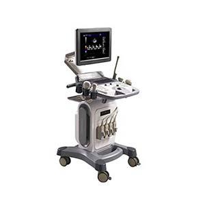
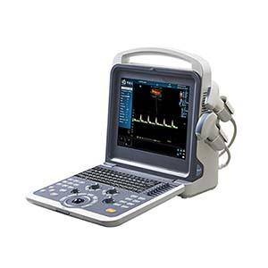
Larger
console with dedicated display
Portability
High;
easy to carry between locations
Generally
less portable
Display
Platform
Compatible
Windows or Android terminal
Usually
an integrated medical display
Imaging
Modes
B,
B/B, B/M, CDFI, PW, PDI
Depends
on system configuration
Frequency
Range
2.5–5.0
MHz
Depends
on probe configuration
Power
Supply
USB-powered
probe
Typically
mains-powered console
Image
Storage
Up
to 2,048 cine frames, plus static images
Depends
on system storage and software
Typical
Applications
Point-of-care
imaging, bedside examinations, emergency screening
General
imaging departments and comprehensive diagnostic workflows
Main
Advantage
Lightweight
design and flexible deployment
Potentially
broader system integration and probe selection
Note: This is a general comparison. Imaging performance,
diagnostic capabilities, and available functions depend on the specific
equipment configuration, software, probe, and clinical application.
Frequently Asked Questions (FAQ)
1. What is the C30C Mini Color Ultrasound Diagnosis System
used for?
The
C30C is designed for portable ultrasound examinations, including abdominal,
gynecological, obstetric, and urological applications, as well as emergency
imaging and point-of-care assessment.
2. Which organs can the C30C examine?
Its
intended applications include the liver, gallbladder, kidneys, spleen,
pancreas, uterus, bladder, and other organs accessible to the configured convex
array probe.
3. Which ultrasound imaging modes are supported?
The
system supports B-mode, B/B-mode, B/M-mode, color Doppler flow imaging (CDFI),
pulsed-wave Doppler (PW), and power Doppler imaging (PDI).
4. What is the operating frequency range?
The
C30C provides six selectable frequencies: 2.5 MHz, 3.0 MHz, 3.5 MHz, 4.0 MHz,
4.5 MHz, and 5.0 MHz.
5. Is the C30C compatible with smartphones or tablets?
The
system supports Windows and Android devices through USB connectivity.
Compatibility depends on the terminal's hardware, operating system, interface,
and the required software.
6. Does the ultrasound probe require a separate battery?
No.
The probe is powered through the USB interface of a compatible terminal. The
specified continuous operating time for the complete system is at least 3 hours
under specified conditions.
7. Can the C30C store ultrasound images and video
sequences?
Yes.
It supports multiple static image formats and can store and play back up to
2,048 image frames. USB transfer and Windows-based DICOM sending are also
supported as specified.
8. Does the system support ultrasound-guided procedures?
It
can support selected ultrasound-guided procedures, including vascular access
and PICC catheter placement, when the probe, imaging modes, and clinical
workflow are appropriate.
9. Can the C30C be used for telemedicine?
Yes.
Images and medical data can be shared through compatible software and connected
terminal devices to support remote consultation workflows. Actual telemedicine
functionality depends on the available software and network infrastructure.