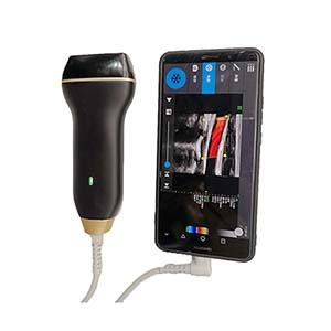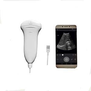
U20L All-Digital Handheld Probe Ultrasonic Diagnostic Instrument

- PRODUCT DETAIL
U20L All-Digital Handheld Probe Ultrasonic Diagnostic
Instrument
Product Introduction
The
U20L All-Digital Handheld Probe Ultrasonic Diagnostic Instrument is a compact,
portable ultrasound imaging system designed for the examination of superficial
tissues, small organs, muscles, and peripheral blood vessels. Featuring digital
beamforming (DBF), real-time dynamic aperture imaging (RDA), dynamic
apodization, and dynamic transmit and receive focusing, it provides versatile
image acquisition and processing for point-of-care clinical applications.
The
U20L supports five imaging modes and a selectable probe frequency range of
5.0–10.0 MHz, making it suitable for superficial ultrasound examinations
requiring higher-frequency imaging. Its adjustable detection depth of 20–120
mm, up to 4× image magnification, and advanced digital image enhancement
functions support detailed assessment of structures such as the thyroid, skin,
muscles, and peripheral vessels.
With
a lightweight handheld design, USB connectivity, multiple image storage
formats, cine-loop playback, and DICOM transmission support, the U20L offers
flexible deployment in hospitals, primary healthcare facilities, emergency
departments, and bedside examination environments. Connection to compatible
computers, tablets, and mobile devices also supports convenient image review
and remote medical collaboration.
Performance Features
• Advanced
Digital Imaging: Integrates digital beamforming (DBF), real-time dynamic
aperture imaging (RDA), dynamic apodization, and dynamic transmit and receive
focusing.
• High-Frequency
Imaging: Provides six selectable frequencies from 5.0 to 10.0 MHz for superficial
tissue, small-organ, muscle, and peripheral vascular examinations.
• Five
Imaging Modes: Supports B, B+B, 4B, B+M, and M modes for flexible image
presentation.
• Adjustable Imaging
Depth: Offers a detection depth range of 20–120 mm to accommodate different
superficial examination requirements.
• Up
to 4× Magnification: Provides eight levels of real-time B-mode
magnification and local enlargement for real-time or frozen images.
• Advanced
Image Processing: Features dynamic digital filtering, image
enhancement, line correlation, frame correlation, linear interpolation,
spatial-temporal filtering, and edge enhancement.
• Flexible Image
Adjustment: Includes an adjustable dynamic range of 0–100 dB,
256-level grayscale conversion, three display-speed settings, and eight-segment
digital time-gain compensation (TGC).
• Cine-Loop Playback: Supports
cyclic storage of up to 512 image frames, with continuous and frame-by-frame
playback.
• Multiple Image Formats: Supports
static image storage in BMP, JPG, PNG, TIF, and DCM formats, together with USB
export and DICOM transmission.
• Multilingual
Interface: Provides Chinese and English menu options, text annotations, measurement
labels, patient information, and probe-position indicators.
• Portable,
Energy-Efficient Design: Uses high-speed integrated circuits and
low-voltage components in a compact handheld housing for flexible clinical
deployment.
• Flexible
Terminal Connectivity: Supports operation with compatible computers,
laptops, tablets, and mobile devices, subject to interface and software
compatibility.
Working Principle
The
U20L uses an electronic array probe to transmit high-frequency ultrasound waves
into superficial tissues and receive echoes reflected by different anatomical
structures. The system processes these echoes to generate diagnostic ultrasound
images.
Digital
beamforming (DBF) coordinates signals received by the probe elements, while
real-time dynamic aperture imaging (RDA), dynamic apodization (DRA), and
dynamic transmit and receive focusing optimize signal processing across the
imaging field. Numerical-control dynamic frequency scanning (DFS) supports
selectable operating frequencies from 5.0 to 10.0 MHz.
The
acquired signals undergo digital filtering, grayscale conversion, image
enhancement, and other processing operations before being displayed. Adjustable
time-gain compensation helps compensate for variations in echo intensity with
depth, while magnification and image-freezing functions facilitate closer
examination of specific structures.
The
instrument also provides measurement tools, cine-loop review, and digital image
storage. Supported USB transfer and DICOM communication facilitate image
archiving and integration with compatible clinical information systems.
Technical Parameters
Parameter
Specification
Product
Model
U20L
Instrument
Type
All-digital
handheld ultrasound diagnostic instrument
Structure
Portable
handheld
Scanning
Method
80-element
electronic array probe; described as convex array in the supplied
specifications
Probe
Interface
USB
Preset
Examination Modes
Small
organs, blood vessels, muscles
Imaging
Modes
B,
B+B, 4B, B+M, M
Probe
Frequency
5.0,
6.0, 7.0, 8.0, 9.0, 10.0 MHz
Detection
Depth
20–120
mm, adjustable
Image
Magnification
8
levels, up to 4×
Dynamic
Range
0–100
dB, adjustable
Grayscale
Conversion
256
levels
Display
Speed
3
adjustable levels
Time-Gain
Compensation
8-segment
digital TGC
Image
Preprocessing
Variable
aperture, dynamic apodization, dynamic digital filtering
Image
Postprocessing
Gamma
correction, line correlation, frame correlation, dot-line correlation, linear
interpolation, spatial-temporal filtering, edge enhancement
Pseudo-Color
Processing
42
options
Cine-Loop
Storage
Up
to 512 frames
Static
Image Formats
BMP,
JPG, PNG, TIF, DCM
Data
Transfer
USB
storage device; DICOM transmission
User
Interface
Chinese/English
menu and input selection
Annotation
Functions
Arrows,
markers, text annotations, patient information, probe-position indicators
Body-Position
Markers
140
Measurement
Functions
Distance,
circumference, area, volume, circular measurement, heart rate, and additional
measurements depending on software version
Compatible
Terminals
Computers,
laptops, tablets, and mobile devices, subject to compatibility
Typical
Applications
Superficial
tissues, thyroid, peripheral blood vessels, muscles, and small organs
Note: The supplied specifications also mention obstetric
measurements, including BPD, GS, CRL, FL, HL, OFD, TTD, and AC. Their
availability should be confirmed for the specific U20L software version and
configuration. The probe geometry should also be verified because the source
identifies the scanning method as an electronic convex array despite the stated
superficial imaging applications.
Application Scenarios
1. Thyroid and Small-Organ Examination
The
U20L's 5–10 MHz frequency selection supports ultrasound examination of the
thyroid and other superficial small organs, helping clinicians assess
anatomical structures and tissue characteristics.
2. Peripheral Vascular Ultrasound
Supports
examination of peripheral blood vessels for clinical assessment and can assist
trained healthcare professionals during selected vascular access procedures.
3. Muscle and Soft-Tissue Imaging
Suitable
for imaging superficial muscles, skin, and soft tissues in clinical departments
requiring portable ultrasound assessment.
4. Bedside and Point-of-Care Diagnostics
Its
handheld design enables convenient deployment in hospital wards, outpatient
departments, and primary healthcare facilities for examinations near the
patient.
5. Emergency and Critical Care
The
portable instrument can support preliminary ultrasound assessments in emergency
departments, intensive care units, ambulances, and field rescue environments,
subject to clinical requirements and device configuration.
6. Ultrasound-Guided Procedures
May
assist trained professionals with vascular localization, anesthesia-related
procedures, PICC catheter placement, and selected image-guided interventions
when appropriate for the probe and intended clinical use.
7. Primary Healthcare and Screening
Suitable
for preliminary examinations in community clinics, physical examination
departments, and other healthcare settings that benefit from portable
diagnostic imaging.
8. Digital Medical Collaboration
USB
export, supported image formats, and DICOM transmission facilitate digital
image management and potential remote consultation through compatible medical
systems.
U20L vs. U20C Handheld Ultrasound Scanner
Comparison Item
U20L
U20C
Primary
Application
Superficial
tissues, small organs, muscles, and peripheral vessels
Abdominal,
obstetric, gynecological, and urological examinations
Probe
Frequency
5.0–10.0
MHz
2.0–5.0
MHz
Adjustable
Detection Depth
20–120
mm
90–240
mm
Imaging
Modes
B,
B+B, 4B, B+M, M
B,
B+B, 4B, B+M, M
Probe
Configuration
Specified
as an 80-element electronic array; geometry requires confirmation
80-element
electronic convex array
Image
Magnification
Up
to 4×
Up
to 4×
Cine-Loop
Storage
Up
to 512 frames
Up
to 512 frames
Image
Storage
BMP,
JPG, PNG, TIF, DCM
BMP,
JPG, PNG, TIF, DCM
Portability
Handheld
Handheld
Typical
Clinical Focus
High-frequency
superficial imaging
Deeper
abdominal and obstetric imaging
The U20L's higher-frequency range is generally suited to
superficial structures, while the U20C's lower-frequency range supports greater
penetration. Actual performance depends on probe design, tissue
characteristics, and device configuration.
Frequently Asked Questions (FAQ)
Q1: What is the U20L handheld ultrasound scanner used for?
The
U20L is designed for ultrasound examinations of superficial tissues, small
organs, muscles, and peripheral blood vessels, including the thyroid and skin
surface.
Q2: What frequencies does the U20L support?
It
provides six selectable frequencies: 5.0, 6.0, 7.0, 8.0, 9.0, and 10.0 MHz.
Q3: What is the imaging depth of the U20L?
The
specified detection depth is adjustable from 20 to 120 mm, supporting
examinations of structures at different superficial tissue depths.
Q4: Which imaging modes are available?
The
U20L supports B, B+B, 4B, B+M, and M imaging modes.
Q5: Can the U20L enlarge ultrasound images?
Yes.
It provides eight levels of real-time B-mode magnification, with a maximum
magnification of 4×. Local enlargement is also available for real-time and
frozen images.
Q6: Does the U20L support image storage and playback?
Yes.
It supports cine-loop storage of up to 512 frames, continuous playback, and
frame-by-frame review. Static images can be saved in BMP, JPG, PNG, TIF, and
DCM formats.
Q7: Can the U20L connect to a computer or mobile device?
The
supplied specifications indicate support for compatible desktop computers,
laptops, tablets, and mobile devices. Actual compatibility depends on the USB
interface, operating system, and required software.
Q8: Can the U20L be used for vascular access guidance?
It
may support vascular localization and selected ultrasound-guided procedures
when the probe and system configuration are suitable. Procedures should be
performed by appropriately trained healthcare professionals.
Q9: What is the difference between the U20L and U20C?
The
U20L operates at 5–10 MHz and is intended primarily for superficial tissue and
small-organ imaging. The U20C operates at 2–5 MHz and is oriented toward
abdominal and obstetric applications requiring greater imaging depth.

NEXT:CU-500 Online Total Organic Carbon (TOC) Analyzer