K10 Full Digital Color Doppler Ultrasound Diagnostic System
Product Introduction
The
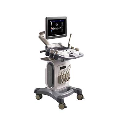
K10 Full Digital Color Doppler Ultrasound Diagnostic System is a versatile
medical imaging system designed for diagnostic examinations across multiple
clinical specialties. Featuring a 15-inch LED monitor, a flexible adjustable
arm, a rotatable operation panel, and four active probe connectors, the K10
supports efficient workflows and flexible probe selection.
Its
advanced imaging functions include directional Doppler flow imaging, tissue
harmonic imaging, speckle reduction, pulse wave Doppler, dynamic focusing, and
Triplex mode. Compatible with multiple transducer types, including convex,
linear, micro-convex, phased-array, transvaginal, and optional 4D volume
probes, the K10 supports examinations in abdominal imaging, obstetrics,
gynecology, pediatrics, vascular assessment, musculoskeletal imaging, and
superficial organ evaluation.
Performance Features
• Full
digital color Doppler imaging: Supports grayscale, color flow, and
spectral Doppler imaging modes.
• 15-inch
LED monitor: Provides a clear display for diagnostic image observation.
• Four active probe
connectors: Enables convenient connection and switching among
compatible transducers.
• Advanced image
optimization: Includes speckle reduction, tissue harmonic imaging,
dynamic apodization, and dynamic aperture.
• Multiple Doppler modes: Supports
directional Doppler flow imaging and pulse wave Doppler, with continuous wave
Doppler available as an option.
• Flexible
focusing system: Features continuous dynamic focusing, 1–16 selectable
transmit focus settings, acoustic lens focus, and 1–4 focus options.
• 10-step
real-time zoom: Offers magnification from ×1.0 to ×10.0 with selectable
zoom positions.
• Cine memory and
storage: Supports B-mode and M-mode cine memory with a 500GB hard drive.
• Customizable
examination settings: Allows users to edit examination types and
save user-defined items.
• Flexible
clinical configuration: Supports multiple transducer types and
optional imaging functions, including panoramic and trapezoid imaging.
Working Principle
The
K10 ultrasound diagnostic system uses piezoelectric transducers to transmit
high-frequency ultrasound waves into the body and receive echoes reflected by
different tissues and structures.
The
system processes the returned signals to generate grayscale images of
anatomical structures. Color Doppler imaging uses frequency shifts caused by
moving blood cells to display blood-flow direction and relative velocity, while
spectral Doppler modes provide flow-related waveform information.
Advanced
image-processing functions, including tissue harmonic imaging, speckle
reduction, dynamic aperture, and dynamic focusing, help optimize image quality.
Optional 4D volume imaging supports three-dimensional volume acquisition with
time-dependent visualization when used with a compatible probe and software
configuration.
Technical Parameters
1. System Configuration
|
Parameter
|
Specification
|
|
Model
|
K10
|
|
Product
Type
|
Full
Digital Color Doppler Ultrasound Diagnostic System
|
|
Monitor
|
15-inch
LED
|
|
Operation
Panel
|
Rotatable
|
|
Support
Arm
|
Flexible
free arm
|
|
Keyboard
|
Backlit
keyboard
|
|
TGC
|
8TGC
|
|
Active
Probe Connectors
|
4
|
|
Probe
Holders
|
6
|
|
Casters
|
4,
with brakes
|
|
Hard
Drive
|
500GB
|
2. Compatible Transducers
|
Probe Type
|
Nominal Frequency
|
Available Frequency Settings
|
|
Convex
Probe
|
3.5
MHz
|
2.0
/ 3.0 / 3.5 / 4.0 / 5.5 MHz
|
|
Linear
Probe
|
7.5
MHz
|
6.0
/ 6.5 / 7.5 / 10.0 / 12.0 MHz
|
|
Micro-Convex
Probe
|
3.5
MHz
|
2.0
/ 2.5 / 3.5 / 4.5 / 5.0 MHz
|
|
Transvaginal
Probe
|
6.5
MHz
|
5.0
/ 6.0 / 6.5 / 7.5 / 9.0 MHz
|
|
Phased-Array
Probe
|
3.5
MHz
|
2.1
/ 3.0 / 3.5 / 4.0 / 5.0 MHz
|
|
4D
Volume Probe
|
3.5
MHz nominal
|
2.0
/ 3.0 / 3.5 / 4.0 / 5.5 MHz
|
Probe availability depends on the selected system
configuration.
3. Imaging Functions and Display Modes
|
Parameter
|
Specification
|
|
Image
Optimization
|
Supported
|
|
Speckle
Reduction
|
Supported
|
|
Tissue
Harmonic Imaging
|
Supported
|
|
Dynamic
Apodization
|
Supported
|
|
Dynamic
Aperture
|
Supported
|
|
Directional
Doppler Flow Imaging
|
Supported
|
|
Pulse
Wave Doppler (PW)
|
Supported
|
|
Continuous
Wave Doppler (CW)
|
Optional
|
|
Panoramic
Imaging
|
Optional
|
|
Trapezoid
Imaging
|
Optional
|
|
TSF
|
Supported
|
|
HPRF
|
Supported
|
|
Triplex
Mode
|
Supported
|
|
Display
Modes
|
B,
B/B, 4B, B/M, M, B/B, B/C/D, B/D, PW, B/PW, CF, CW, 4D
|
|
Real-Time
Zoom
|
×1.0
to ×10.0, 10 steps
|
|
Zoom
Position
|
Selectable
|
|
Transmit
Focus
|
1–16
selectable settings
|
|
Acoustic
Lens Focus
|
Supported
|
|
Focus
Options
|
1,
2, 3, 4
|
4. Connectivity and Storage
|
Parameter
|
Specification
|
|
Video
Output
|
Available
|
|
VGA
Output
|
Available
|
|
RS-232
|
Available
|
|
USB
Ports
|
2
|
|
DICOM
|
DICOM
3.0
|
|
Cine
Memory
|
Supported
|
|
B-Mode
Memory
|
Supported
|
|
M-Mode
Memory
|
Supported
|
|
Internal
Storage
|
500GB
hard drive
|
5. Physical Specifications
|
Parameter
|
Specification
|
|
Net
Dimensions (L × W × H)
|
750
× 600 × 1380 mm
|
|
Gross
Dimensions (L × W × H)
|
780
× 690 × 1380 mm
|
|
Net
Weight
|
65
kg
|
|
Gross
Weight
|
105
kg
|
6. Power and Operating Conditions
|
Parameter
|
Specification
|
|
Power
Supply Voltage
|
AC
100–242 V
|
|
Frequency
|
50
Hz ± 1 Hz
|
|
Rated
Power
|
≤250
VA
|
|
Ambient
Temperature
|
0°C
to +40°C
|
|
Relative
Humidity
|
38%–85%
|
|
Atmospheric
Pressure
|
700–1060
hPa
|
7. Standard and Optional Accessories
|
Category
|
Accessories
|
|
Standard
Accessories
|
Power
cable, operation manual, fuse, recovery system disc, DVD-ROM
|
|
Optional
Accessories
|
Black-and-white
or color video printer, LaserJet or inkjet printer, biopsy guide, foot switch
|
Application Scenarios
• Abdominal
ultrasound: Examination of abdominal organs and related anatomical
structures.
• Obstetrics and
gynecology: Pregnancy-related ultrasound examinations and
gynecological imaging using compatible probes.
• Pediatric ultrasound: Diagnostic
imaging for pediatric patients with suitable transducers.
• Vascular ultrasound: Color and
spectral Doppler assessment of blood flow.
• Superficial
organ imaging: Examination of superficial tissues and small structures
using high-frequency linear probes.
• Musculoskeletal
imaging: Evaluation of muscles, tendons, joints, and related structures.
• Orthopedic
applications: Ultrasound assessment of selected musculoskeletal
conditions.
• Small-parts imaging: Examination
of superficial anatomical structures using compatible high-frequency
transducers.
K10 Color Doppler Ultrasound System Comparison
|
Feature
|
K10 System
|
Basic B-Mode Ultrasound System
|
|
Imaging
Technology
|
Full
digital color Doppler
|
Primarily
grayscale imaging
|
|
Blood-Flow
Assessment
|
Color
and spectral Doppler modes
|
Generally
unavailable or limited
|
|
Monitor
|
15-inch
LED
|
Varies
by model
|
|
Probe
Connections
|
4
active connectors
|
Varies
by model
|
|
Transducer
Selection
|
Convex,
linear, micro-convex, phased-array, transvaginal, optional 4D
|
Depends
on configuration
|
|
Advanced
Imaging
|
Harmonic
imaging, speckle reduction, dynamic focusing
|
Depends
on model
|
|
Zoom
|
Real-time,
up to ×10
|
Depends
on model
|
|
Data
Connectivity
|
USB,
VGA, video, RS-232, DICOM 3.0
|
Depends
on model
|
|
Storage
|
500GB
hard drive
|
Depends
on model
|
|
Clinical
Applications
|
Abdominal,
obstetric, gynecological, vascular, musculoskeletal, and other examinations
|
Depends
on imaging functions and probes
|
Frequently Asked Questions
1. What is the K10 Color Doppler Ultrasound System used
for?
The
K10 is designed for diagnostic ultrasound examinations in abdominal imaging,
obstetrics, gynecology, pediatrics, vascular assessment, superficial organ
imaging, orthopedics, and musculoskeletal applications.
2. What types of probes are compatible with the K10?
Supported
probe types include convex, linear, micro-convex, transvaginal, phased-array,
and 4D volume probes. Specific probes and frequencies depend on the selected
configuration.
3. How many probes can be connected simultaneously?
The
system has four active probe connectors, allowing up to four compatible probes
to be connected at the same time.
4. Does the K10 support 4D ultrasound imaging?
The
system lists 4D display mode and a 4D volume probe. Confirm that the required
4D probe, software, and licensing are included in the chosen configuration.
5. Does the K10 support vascular Doppler examinations?
Yes.
Directional color Doppler flow imaging and pulse wave Doppler are supported.
Continuous wave Doppler is available as an option.
6. What image optimization functions are available?
The
K10 includes speckle reduction, tissue harmonic imaging, dynamic apodization,
dynamic aperture, and continuous dynamic focusing.
7. Can the system store and export examination data?
The
K10 has a 500GB hard drive, cine memory, two USB ports, video output, VGA
output, RS-232, and DICOM 3.0 connectivity. Actual data transfer and
integration depend on configuration and compatible systems.
8. What is the maximum zoom magnification?
The
real-time zoom function provides 10 selectable steps, from ×1.0 to ×10.0, with
selectable zoom positions.
9. What accessories are available?
Standard
accessories include a power cable, operation manual, fuse, recovery system
disc, and DVD-ROM. Optional accessories include video printers, LaserJet or
inkjet printers, a biopsy guide, and a foot switch.