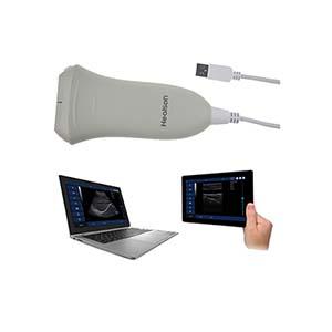
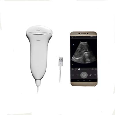
U20C All-Digital Handheld Probe Ultrasonic Diagnostic Instrument
- PRODUCT DETAIL
U20C All-Digital Handheld Probe Ultrasonic Diagnostic Instrument
Product Introduction
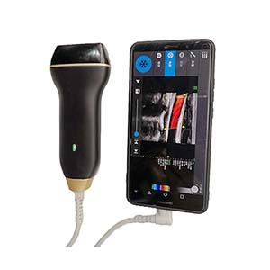
The U20C All-Digital Handheld Probe Ultrasonic Diagnostic Instrument is a compact, portable ultrasound imaging system designed for clinical examinations, emergency diagnostics, and point-of-care applications. It integrates advanced digital beamforming, real-time dynamic aperture imaging, dynamic focusing, digital filtering, and image enhancement technologies to provide versatile ultrasound imaging in a lightweight handheld format.
Equipped with an 80-element electronic convex array probe and five imaging modes, the U20C supports examinations of abdominal organs, the urinary system, and the female reproductive system, as well as obstetric assessments and fetal measurements. Its adjustable imaging depth, multiple image processing functions, and comprehensive measurement capabilities help healthcare professionals perform routine examinations and preliminary assessments.
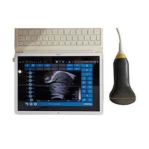
The instrument supports image storage, cine-loop playback, USB data transfer, and DICOM transmission. It can connect to compatible computers, laptops, tablets, and mobile devices, offering flexible deployment in hospitals, primary healthcare facilities, emergency units, ambulances, and remote medical environments.
Performance Features
• Advanced Digital Imaging: Uses digital beamforming (DBF), real-time dynamic aperture imaging (RDA), dynamic sound velocity apodization (DRA), and dynamic transmit and receive focusing for versatile ultrasound imaging.
• Multiple Imaging Modes: Supports B, B+B, 4B, B+M, and M modes to accommodate different clinical examination requirements.
• Wide Frequency Selection: Provides five selectable probe frequencies: 2.0, 3.0, 3.5, 4.0, and 5.0 MHz.
• Adjustable Imaging Depth: Offers a detection depth of at least 190 mm, with adjustable settings from 90 to 240 mm.
• Advanced Image Processing: Includes dynamic digital filtering, image enhancement, grayscale conversion, spatial-temporal filtering, edge enhancement, and 42 pseudo-color processing options.
• Flexible Image Magnification: Features eight levels of real-time B-mode magnification, up to 4×, plus local magnification for real-time and frozen images.
• Comprehensive Measurement Tools: Supports distance, circumference, area, volume, ellipse, heart rate, fetal biometric measurements, estimated delivery date, fetal weight, and amniotic fluid index, depending on the software version.
• Cine-Loop Review: Stores up to 512 image frames for continuous or frame-by-frame playback.
• Multiple Data Formats: Supports static image storage in BMP, JPG, PNG, TIF, and DCM formats, as well as USB export and DICOM transmission.
• Portable and Energy-Efficient Design: Uses high-speed integrated circuits and low-voltage components to provide a compact, lightweight instrument suitable for mobile clinical use.
• Flexible Terminal Connectivity: Supports connection to compatible desktop computers, laptops, tablets, and mobile devices for convenient operation and image review.
• Broad Clinical Applications: Suitable for emergency assessment, bedside examinations, obstetric screening, anesthesia guidance, vascular localization, and other ultrasound-assisted procedures.
Working Principle
The U20C operates by transmitting high-frequency ultrasound waves into the body through an electronic convex array probe and receiving echoes reflected by internal tissues and organs. The system processes these echo signals to reconstruct ultrasound images for clinical interpretation.
Its digital beamforming (DBF) technology coordinates signals from multiple probe elements. Real-time dynamic aperture imaging and dynamic transmit and receive focusing optimize signal acquisition across different imaging depths. Dynamic frequency scanning and eight-segment digital time-gain compensation (TGC) help adjust image acquisition and compensate for differences in echo intensity.
The processed signals undergo digital filtering, grayscale conversion, image enhancement, and other image processing operations before being displayed. Depending on the selected mode, the system presents two-dimensional B-mode images, combined B-mode views, four-panel images, or motion-related M-mode displays.
Measurement tools and cine-loop playback support further examination and review. Digital storage, USB transfer, and DICOM communication facilitate image documentation and integration with compatible medical information workflows.
Technical Parameters
|
Parameter |
Specification |
|
Product Model |
U20C |
|
Instrument Type |
All-digital handheld ultrasound diagnostic instrument |
|
Structure |
Portable handheld type |
|
Scanning Method |
80-element electronic convex array |
|
Probe Interface |
USB |
|
Imaging Modes |
B, B+B, 4B, B+M, M |
|
Probe Frequency |
2.0, 3.0, 3.5, 4.0, 5.0 MHz |
|
Detection Depth |
≥190 mm |
|
Adjustable Depth Range |
90–240 mm |
|
Real-Time Image Magnification |
8 levels, up to 4× |
|
Dynamic Range |
0–100 dB, adjustable |
|
Grayscale Conversion |
256 levels |
|
Display Speed |
3 adjustable levels |
|
Time-Gain Compensation |
8-segment digital TGC |
|
Image Processing |
Dynamic aperture, dynamic apodization, digital filtering, image enhancement, linear interpolation, spatial-temporal filtering |
|
Gamma Correction |
8 types |
|
Pseudo-Color Processing |
42 options |
|
Cine-Loop Storage |
Up to 512 frames |
|
Static Image Formats |
BMP, JPG, PNG, TIF, DCM |
|
Data Transfer |
USB storage device; DICOM transmission |
|
User Interface |
Chinese/English menu and input selection |
|
Annotation Functions |
Arrows, markers, text annotations, body-position indicators |
|
Patient Information |
Patient name, age, gender, hospital name, and related information |
|
Body-Position Markers |
140 |
|
Measurement Functions |
Distance, circumference, area, volume, ellipse, heart rate, and selected obstetric measurements, depending on version |
|
Compatible Terminals |
Computers, laptops, tablets, and mobile devices, subject to compatibility |
|
Typical Applications |
Abdominal, gynecological, obstetric, urological, emergency, and point-of-care examinations |
Note: Detailed measurement functions, terminal compatibility, and DICOM connectivity may vary by software version and configuration. Confirm the final specifications with the supplier before purchase.
Application Scenarios
1. Abdominal Ultrasound Examination
Supports ultrasound assessment of the liver, gallbladder, kidneys, spleen, and pancreas for routine clinical examination and preliminary diagnostic evaluation.
2. Obstetrics and Gynecology
Facilitates obstetric examinations, fetal biometric measurements, pregnancy assessments, uterine imaging, and examinations of the female reproductive system. Available measurements may include BPD, GS, CRL, FL, HL, OFD, TTD, and AC, depending on the software version.
3. Urological Examination
Supports ultrasound assessment of the kidneys and bladder, helping healthcare professionals evaluate relevant anatomical structures.
4. Emergency and Point-of-Care Diagnostics
Its portable design makes the U20C suitable for emergency departments, intensive care units, ambulances, field rescue operations, bedside examinations, and other situations requiring flexible ultrasound access.
5. Primary Healthcare and Screening
Can be used in community medical facilities, hospital wards, physical examination departments, family planning services, and primary healthcare settings for preliminary screening and routine examinations.
6. Ultrasound-Guided Procedures
May assist trained healthcare professionals with ultrasound-guided anesthesia, vascular localization, PICC catheter placement, and other image-guided interventions, subject to the probe, clinical protocol, and intended-use configuration.
7. Remote Medical Collaboration
Digital image storage and supported data transmission enable ultrasound images and examination data to be shared with compatible systems for remote consultation and telemedicine workflows.
U20C vs. Conventional Ultrasound Diagnostic Systems
|
Comparison Item |
U20C Handheld Ultrasound |
Conventional Cart-Based Ultrasound |
|
Portability |
Compact handheld design |
Typically larger and less mobile |
|
Deployment |
Suitable for bedside, emergency, and mobile examinations |
Commonly used in dedicated examination rooms |
|
Imaging Modes |
B, B+B, 4B, B+M, M |
Varies by system and configuration |
|
Probe Technology |
80-element electronic convex array |
Varies by probe and system |
|
Image Processing |
Digital beamforming, dynamic focusing, and digital enhancement |
Depends on the imaging platform |
|
Data Management |
Supports multiple image formats, USB export, and DICOM transmission |
Depends on system configuration |
|
Terminal Flexibility |
Compatible with selected computers and mobile devices |
Often uses an integrated display and control console |
|
Typical Use |
Point-of-care examinations and preliminary assessment |
Routine and specialized diagnostic imaging |
Actual imaging performance, clinical capabilities, and workflow features depend on the specific equipment configuration. This comparison describes general design differences rather than a claim of equivalent diagnostic performance.
Frequently Asked Questions (FAQ)
Q1: What is the U20C handheld ultrasound diagnostic instrument used for?
The U20C is designed for ultrasound examinations involving abdominal organs, the urinary system, obstetrics, and gynecology. It can also support emergency assessment, bedside examinations, and selected ultrasound-guided procedures.
Q2: Which imaging modes does the U20C support?
It supports five modes: B, B+B, 4B, B+M, and M, providing different image display options for clinical examination.
Q3: What is the operating frequency range of the U20C probe?
The instrument provides five selectable frequencies: 2.0, 3.0, 3.5, 4.0, and 5.0 MHz.
Q4: What is the maximum detection depth?
The specified detection depth is at least 190 mm, with an adjustable depth range of 90–240 mm.
Q5: Can the U20C perform obstetric measurements?
Yes. Depending on the software version, it can support fetal biometric measurements such as BPD, GS, CRL, FL, HL, OFD, TTD, and AC, as well as estimated delivery date, fetal weight, and amniotic fluid index calculations.
Q6: Does the instrument support image storage and playback?
Yes. It can store up to 512 image frames for cine-loop playback and supports static image formats including BMP, JPG, PNG, TIF, and DCM.
Q7: Can ultrasound images be transferred to a computer or USB drive?
The U20C supports USB storage-device export and connection to compatible computer terminals. DICOM transmission is also specified, subject to system and software compatibility.
Q8: Is the U20C suitable for emergency and mobile medical applications?
Yes. Its handheld construction and flexible terminal connectivity make it suitable for emergency departments, intensive care units, ambulances, field rescue, bedside examinations, and primary healthcare settings.
Q9: Can the U20C replace a conventional cart-based ultrasound system?
Not necessarily. The U20C is intended for portable examinations and selected clinical applications. Whether it meets a facility's requirements depends on diagnostic needs, available probes, software functions, image quality, and regulatory indications.
NEXT:U20L All-Digital Handheld Probe Ultrasonic Diagnostic Instrument