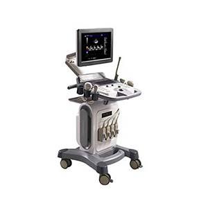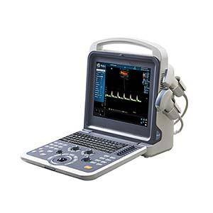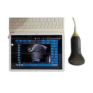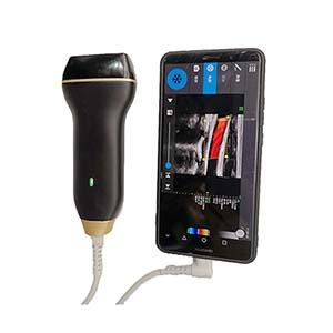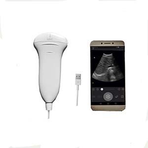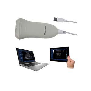
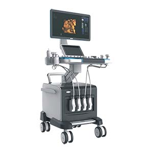
MDK-9900 Full Digital Color Doppler Ultrasound Diagnostic System
- PRODUCT DETAIL
MDK-9900 Full Digital Color Doppler Ultrasound Diagnostic System
Product Introduction
The MDK-9900 Full Digital Color Doppler Ultrasound Diagnostic System is a versatile medical ultrasound imaging platform designed for diagnostic examinations across multiple clinical departments. It supports two-dimensional grayscale imaging, color Doppler flow imaging, color power Doppler, directional energy Doppler, pulsed-wave Doppler (PWD), continuous-wave Doppler (CW), M-mode, harmonic imaging, composite imaging, and variable-aperture imaging.
Equipped with a 21.5-inch widescreen high-resolution color LCD monitor, a 12.1-inch touchscreen, and a 50 mm trackball, the MDK-9900 provides an integrated interface for image acquisition, measurement, analysis, and patient record management. Its probe options cover abdominal, superficial, cardiac, and intracavitary examinations, with optional 4D imaging available.
The system supports DICOM 3.0 connectivity, HDMI, VGA video output, USB storage, and wireless transmission as specified. It can store and replay static and dynamic ultrasound images on its internal hard disk or compatible USB storage devices.
Performance Features
1. Comprehensive imaging modes: Supports 2D grayscale, color Doppler, color power Doppler, directional energy Doppler, PWD, CW, M-mode, harmonic, composite, and variable-aperture imaging.
2. Large widescreen display: Features a 21.5-inch high-resolution LCD monitor with flicker-free progressive scanning and adjustable viewing position.
3. Interactive touchscreen: A 12.1-inch widescreen touchscreen supports convenient system operation.
4. Multi-probe compatibility: Optional abdominal, high-frequency linear, cardiac, and intracavitary probes support a wide range of examinations.
5. Adjustable imaging frequency: Provides at least six frequency conversion options in 2D mode, with selectable Doppler frequencies.
6. Advanced grayscale imaging: Uses digital beam forming, full-range dynamic focusing, dynamic aperture, dynamic apodization, and sidelobe compression technologies.
7. Image enhancement: Speckle-noise purification and noise-reduction technologies help improve image presentation.
8. Comprehensive Doppler analysis: Supports Doppler automatic envelope detection and measurement, color flow adjustment, and multiple flow display modes.
9. Flexible scanning controls: Offers adjustable scan depth, fan scan angle, gain, time-gain compensation (TGC), and acoustic output power.
10. Integrated measurement software: Includes general measurements, obstetric calculations, cardiac function analysis, vascular flow analysis, color Doppler measurements, and urology measurements.
11. Image and patient data management: Supports static and dynamic image storage, playback, archiving, and retrieval.
12. DICOM 3.0 connectivity: Supports connection with compatible medical imaging and communication systems.
13. Multiple output and storage options: Provides VGA, HDMI, USB interfaces, hard-disk storage, and supported wireless transmission.
14. Optional 4D imaging: Optional 4D capability can extend the system's imaging functions when supported by the required configuration.
Working Principle
The MDK-9900 uses digital ultrasound transmission and reception to generate diagnostic images. The transducer emits ultrasound pulses into the body and receives echoes reflected by tissues and anatomical structures. The system processes these signals through digital beam forming, dynamic focusing, gain control, and image enhancement to produce two-dimensional grayscale images.
For Doppler examinations, the system analyzes frequency shifts in echoes from moving blood cells to assess blood-flow direction and velocity. Color Doppler and energy-based modes display flow information over the grayscale image, while spectral Doppler presents flow-related signals for further analysis. M-mode supports motion assessment, and harmonic and composite imaging provide additional image-processing options.
The system's measurement software processes acquired images and Doppler data to support general, obstetric, cardiac, vascular, and urological assessments. Static and dynamic images can be saved and reviewed through the system's image-management functions.
Technical Parameters
1. General System Specifications
|
Parameter |
Specification |
|
Model |
MDK-9900 |
|
System type |
Full digital color Doppler ultrasound diagnostic system |
|
Operating system |
Windows 10 |
|
Main monitor |
21.5-inch widescreen high-resolution color LCD |
|
Monitor characteristics |
Flicker-free, progressive scanning; adjustable viewing position |
|
Touchscreen |
12.1-inch widescreen |
|
Trackball |
50 mm |
|
Probe connectors |
4 or more zero-insertion-force metal-body probe interfaces |
|
Probe interface specification |
256-core probe interface, as specified |
|
Imaging modes |
2D, color Doppler, color power Doppler, directional energy Doppler, PWD, CW, M-mode, harmonic, composite, variable aperture |
|
Maximum system scanning depth |
34 cm |
|
Dynamic range |
20–280 dB, continuously adjustable |
|
Physical receiving channels |
64 or more |
|
A/D conversion |
12 bit or higher |
|
TGC adjustment |
8 segments or more |
|
Maximum grayscale cine playback |
600 images or more |
|
Acoustic output |
Visual acoustic output power display and adjustment |
|
Optional function |
4D imaging |
2. Probe Specifications
|
Probe Type |
Frequency / Feature |
Additional Specification |
|
Abdominal convex probe |
2.5–5.0 MHz |
Maximum detection depth ≥19 cm |
|
High-frequency linear probe |
6.0, 7.5, 8.5, 10, 11, and 12 MHz |
Supports 128- and 256-element array probes |
|
Intracavitary probe |
Frequency depends on configuration |
Scanning angle: 151° |
|
Cardiac probe |
Optional |
Frequency and detailed specifications depend on configuration |
|
Probe frequency adjustment |
At least 6 frequency conversion options in 2D mode |
Different frequencies can be selected for Doppler |
|
B/D dual-use modes |
Linear array: B/PWD; convex array: B/PWD; fan scan: B/PWD |
Depends on compatible probe |
|
Puncture guide |
Optional |
Probe-compatible puncture guide device |
3. Two-Dimensional Grayscale Imaging
|
Parameter |
Specification |
|
Maximum scanning depth |
34 cm |
|
Dynamic range |
20–280 dB, continuously adjustable |
|
Receiving channels |
≥64 physical channels |
|
Beam-forming technology |
Digital beam forming |
|
Focusing |
Full-range dynamic focusing; adjustable focus position throughout the imaging zone |
|
Aperture and apodization |
Digital dynamic variable aperture and dynamic apodization |
|
Sidelobe control |
Dynamic sidelobe compression |
|
Transmission |
Optimized emission waveform |
|
Image enhancement |
Speckle-noise purification and noise reduction |
|
Gain control |
Independent B-mode and M-mode gain adjustment |
|
TGC |
≥8 segments |
|
Fan scan |
Adjustable fan scan angle |
|
Cine playback |
≥600 grayscale images |
|
Diagnostic presets |
Examination mode selection by anatomical region |
|
Acoustic output |
Visually adjustable |
4. Spectral Doppler Specifications
|
Parameter |
Specification |
|
Pulsed-wave Doppler |
PWD |
|
Doppler frequency |
Two frequency groups each for linear-array PWD and convex-array PWD |
|
Envelope processing |
Automatic Doppler envelope detection and measurement |
|
Color filter |
Automatic and manual adjustment |
|
Wall-filter optimization |
Automatic optimization in response to pulse repetition frequency adjustment |
|
Color baseline |
±9 adjustment levels |
|
Display reversal |
Left/right and up/down reversal |
|
Refresh control |
B-refresh |
|
Playback |
Automatic cine playback |
|
Time-domain filtering |
Real-time color blood-flow adjustment |
5 . Color Doppler Specifications
|
Parameter |
Specification |
|
Doppler gain |
Continuously adjustable |
|
Display modes |
Velocity-variance, energy, velocity, and independent variance displays |
|
Combined display |
2D image, spectral Doppler, and color blood-flow imaging displayed simultaneously |
|
Linear scan region of interest |
−15° to +15° |
|
Flow adjustment |
Real-time color blood-flow adjustment |
|
Color filtering |
Automatic and manual technologies |
6. Measurement, Connectivity, and Data Management
|
Parameter |
Specification |
|
General measurements |
Distance, area, circumference, volume, angle, time, heart rate, flow velocity, flow ratios, and other measurements |
|
Obstetric software |
Obstetric measurements with fetal-weight calculation formula |
|
Cardiac analysis |
Cardiac function measurement and analysis; editable cardiac reports |
|
Vascular analysis |
Vascular blood-flow measurement and analysis |
|
Color Doppler analysis |
Blood-flow measurement and analysis |
|
Urology analysis |
Urological measurement and analysis |
|
Annotation tools |
Chinese and English comments; insertion, deletion, editing, and saving |
|
Video output |
VGA, with RGB conversion as specified |
|
Digital output |
HDMI |
|
USB |
3 USB interfaces |
|
Wireless transmission |
Supported as specified; configuration should be confirmed |
|
Medical imaging connectivity |
DICOM 3.0 |
|
Storage media |
Internal hard disk and USB removable storage |
|
Image management |
Storage, management, playback, archiving, and retrieval of static and dynamic images |
|
4D imaging |
Optional |
Application Scenarios
The MDK-9900 is designed for use in medical institutions requiring a multi-application ultrasound diagnostic system.
• Abdominal imaging: Ultrasound assessment of abdominal organs and related structures.
• Cardiology: Cardiac imaging and functional analysis using compatible cardiac probes and imaging modes.
• Obstetrics and gynecology: Obstetric measurements, fetal-weight estimation, and intracavitary examinations with compatible probes.
• Urology: Urological imaging and measurement analysis.
• Pediatrics and neonatal care: Ultrasound examinations for pediatric and newborn patients using suitable probes and protocols.
• Vascular diagnostics: Color and spectral Doppler assessment of blood flow.
• Superficial structures: High-frequency linear-probe imaging for superficial tissues.
• Breast and thyroid examinations: Ultrasound evaluation of superficial organs.
• Intracavitary examinations: Imaging using compatible intracavitary probes.
• Brain-related examinations: Applications where the appropriate probe, examination protocol, and clinical indication support the intended study.
Actual clinical applications depend on the selected probe, system configuration, approved indications, and local medical-device requirements.
MDK-9900 vs. Conventional Ultrasound Systems
|
Feature |
MDK-9900 |
Conventional Basic Ultrasound System |
|
Imaging modes |
Multiple grayscale, Doppler, M-mode, harmonic, and composite modes |
Often offers a more limited selection, depending on model |
|
Color flow imaging |
Color Doppler, color power Doppler, and directional energy Doppler |
May offer fewer flow-imaging modes |
|
Spectral Doppler |
PWD and specified CW imaging capability |
Frequently configuration-dependent |
|
Display |
21.5-inch LCD plus 12.1-inch touchscreen |
Display size and control interface vary |
|
Probe support |
Abdominal, linear, cardiac, and intracavitary options |
Probe selection varies by system |
|
Image enhancement |
Digital beam forming, speckle-noise purification, and noise reduction |
Image-processing features vary |
|
Measurement software |
General, obstetric, cardiac, vascular, color Doppler, and urology functions |
May provide fewer specialized packages |
|
Image storage |
Internal hard disk and USB storage for static and dynamic images |
Storage options vary |
|
Medical network integration |
DICOM 3.0 interface |
May require optional hardware or software |
|
4D imaging |
Optional |
Depends on system model and configuration |
Frequently Asked Questions (FAQ)
Q1: What is the MDK-9900 Full Digital Color Doppler Ultrasound System used for?
It is designed for a broad range of ultrasound examinations, including abdominal, cardiac, obstetric, gynecological, urological, vascular, breast, thyroid, pediatric, neonatal, and intracavitary applications, subject to compatible probes and clinical indications.
Q2: Which imaging modes does the MDK-9900 support?
Its specified modes include 2D grayscale, color Doppler, color power Doppler, directional energy Doppler, pulsed-wave Doppler, continuous-wave Doppler, M-mode, harmonic imaging, composite imaging, and variable-aperture imaging.
Q3: What display configuration does the system use?
The system has a 21.5-inch widescreen high-resolution color LCD monitor and a separate 12.1-inch widescreen touchscreen. A 50 mm trackball is also specified.
Q4: What probes are available for the MDK-9900?
Optional probe types include abdominal, high-frequency linear, cardiac, and intracavitary probes. The abdominal probe is specified at 2.5–5.0 MHz, while the high-frequency linear probe offers selectable frequencies from 6.0 to 12.0 MHz.
Q5: What is the maximum scanning depth?
The system's maximum scanning depth is specified as 34 cm. The abdominal probe's maximum detection depth is separately listed as 19 cm or more.
Q6: Does the MDK-9900 support obstetric and cardiac measurements?
Yes. The specified measurement software includes obstetric calculations with a fetal-weight formula, cardiac function analysis, editable cardiac reports, vascular flow analysis, general measurements, and urology measurements.
Q7: Can ultrasound images and patient records be stored and reviewed?
Yes. The system supports storage and playback of static and dynamic images on the internal hard disk and compatible USB storage. It also provides image archiving, management, and retrieval functions.
Q8: Does the MDK-9900 support DICOM?
Yes. A DICOM 3.0 interface is specified for communication with compatible medical imaging systems. Integration depends on the receiving system and network configuration.
Q9: Is 4D ultrasound included as standard?
No. The product specifications list 4D imaging as an optional function. Confirm the required probes, software, and hardware configuration with the supplier when ordering.
NEXT:YDMKY Miniature Pore Water Pressure Gauge